
Protocol for Imaging Seed Features
Protocol for Imaging Seed Morphological Features
Do you know?
There are over 200,000 plant species distributed around the world which produce seed and that seed production is the main method of plant propagation. Seed is also an agent for long distance plant dispersal and weed spread. Seeds may be dispersed naturally, and intentionally or unintentionally through commercial trade.
Here, seed is defined as any propagules, disseminules or diaspores, a part of a plant that serves to propagate it. This includes true botanical seeds, dry fruits, fruit fragments, and vegetative reproduction parts such as bulbs.
Determining plant taxonomic identity mainly using seed morphology, known as seed identification, is a highly specialized area of plant taxonomy. Seed identification is an important diagnostic tool for many trade-related systems such as seed certification, phytosanitary certification, seed and grain grading, and commodity labeling. It is also useful in medical, criminal, and veterinary applications, in food science, and for archaeological and anthropological studies.
Why do we image seeds?
Images of seed morphological features are used as virtual specimen references for seed identification and for the production of training materials and reference resources such as identification fact sheets and web-based identification tools. Because of the rich diversity of flowering plant species, only a collective or collaborative approach will achieve the goal to build a large-scale, global reference database. Therefore, the standardization of imaging seed morphological features is important for building such a comprehensive database from various sources over the long term.
Scope:
To describe a standard method or procedure for taking digital images of seeds and their morphological features.
Purpose:
-
To provide standard procedures and the minimum requirements for taking seed images and documenting seed morphological features.
-
To provide a standard format or form for images that are functional for diagnostic purposes, have consistent appearance over time, and display professional quality for a seed image database as the virtual reference for seed morphology and identification.
General Rules
Seed Materials or Specimens Used
Seed materials or specimens must be verified for their taxonomic identity prior to images being taken. The accuracy of images and their associated information is the sole responsibility of the authors who provided the materials. It is recommended that the taxonomic identity be verified by personnel who have seed identification or plant taxonomy expertise.
The selection of seeds for imaging should:
- Represent intended seed morphological features.
- Be sufficient for seed or feature variation range of the species, including the dimorphic or heteromorphic forms present in certain species. For example,
 |
 |
 |
| Variation in size and colour of Ambrosia trifida (Asteraceae) fruits | Variation in size and colour of Acanthospermum hispidum (Asteraceae) achenes | Dimorphic achenes of Centaurea solstitialis (Asteraceae) |
- Represent mature seeds, unless the particular intention is to show the development of maturity, or the morphological variation of maturity.
Images should:
-
Clearly display the morphological features intended
-
Illustrate the range of variation within the species
-
Must be labeled with scientific name and serial number for ease of posting; such as Zea mays-01, Zea mays-02….and so on. Additional information can be added for your organization, such as Zea mays-01-USDA
-
Copyright of an organization or an author can be imbedded on the images
Operation and Recommendations
Images are recommended to be taken with the following measures to be more effective:
-
Have an imaging plan to cover all contents listed in the 'Capturing Seed Morphological Features' section below with a minimum number of images
-
Select seeds or fruits representing features and variations to be taken
-
Provide guidance and advice to the photographer on images taken, if the specialist is not the one taking images for the intended or best presentation
-
Ensure a standard image background is used and appropriately applied to the features and variations intended. The background should not detract in any way from the seed image, not mask any delicate structures or subtle features and focus on the true colour of the seeds. Recommended background for seed imaging is white, black, or grey, and calibrated by image editing software when necessary for the same colour shade.
-
All images must have an accurate scale in relation to the imaged seeds. The authors are fully responsible for the accuracy of scale labels with measures such as routine calibration with a standard reference ruler (usually microscope rulers)
-
It is recommended to review the final images for accuracy, intended presentation, and correct image labelling including scales.
-
In order to present the detailed features of seeds, high-powered in-depth focus, imaging scanning, or stacking is recommended for imaging equipment. Please see recommended image equipment suppliers.
Capturing Seed Morphological Features
To present seed morphological features for identifying a species in full coverage, seed images of a species must ensure features and variations be taken, if materials are available. To display the full range of features and variations within a species, typically four types of images are recommended:
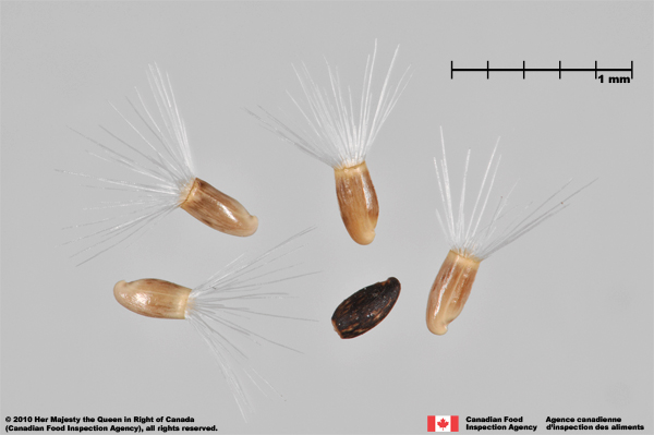
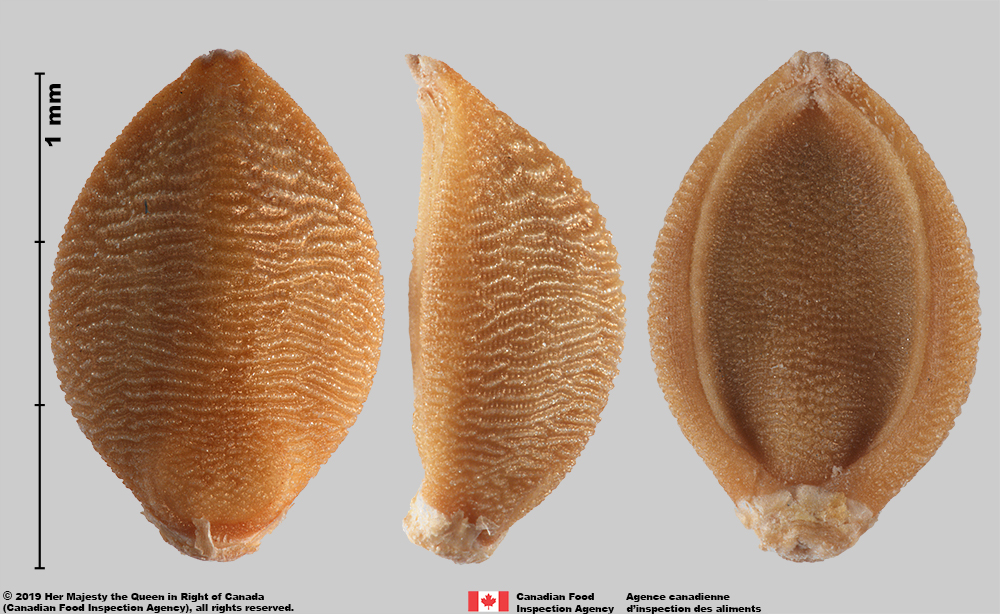
Image Type I: Image(s) of the morphology of the single seed or a dispersal unit: a complete display of the morphological features of an individual seed using:
- A single seed. For example,
Silene latifolia (Caryophyllaceae) seed
- Multiple seeds can be used for completing the morphological image of a single seed. When a species has two or more unique sides, photographing two seeds, each with a different side up may be necessary. For example,
|
|
|
|
A spikelet of Bothriochloa barbinodis (Poaceae) |
A floret of Paspalum laeve (Poaceae) |
 |
 |
|
A floret of Setaria pumila subsp. pumila (Poaceae) |
A seed of Asclepias speciosa (Apocynaceae) |
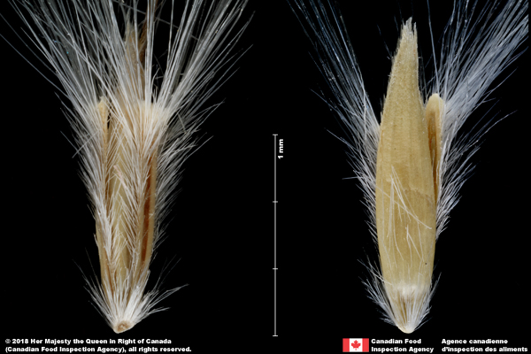
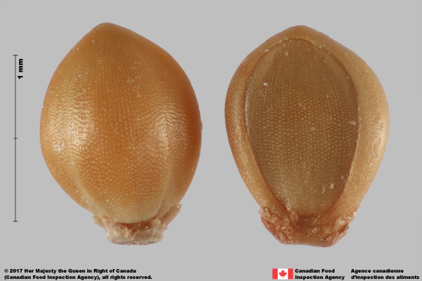
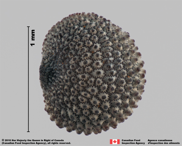
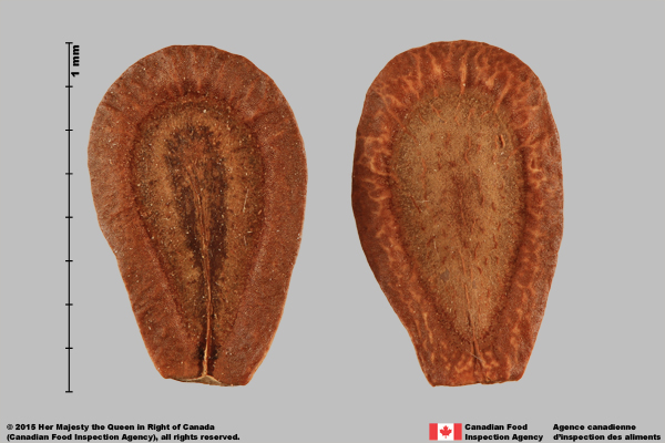
Image Type II: Close-ups for special or unique identification features:
|
|
|
|
|
|
|
| Cross-section of Rumex sp. (Polygonaceae) achene | Longitudinal section of Proboscidea louisianica (Martyniaceae) seed |
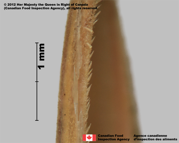
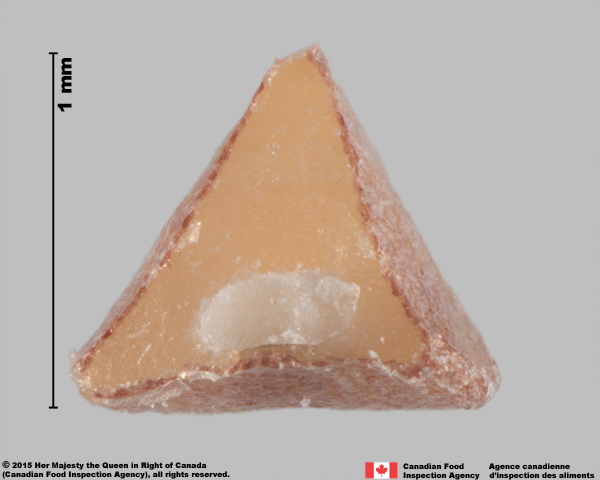
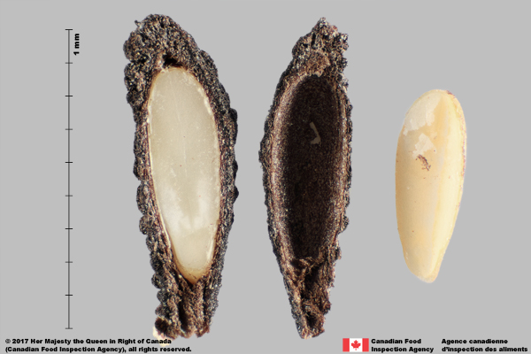
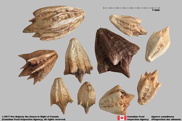
Image Type III: Image(s) of multiple forms of dispersal units and their associated parts: such as spikelet, floret or caryopsis for grass species and achene, seed, remnant floral parts, or bur for aster species.
|
|
|
|
|
|
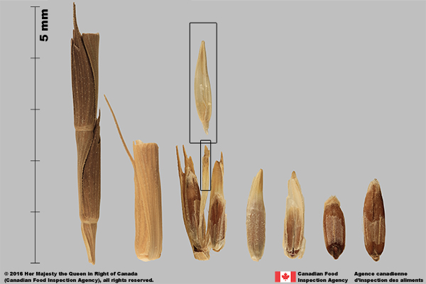
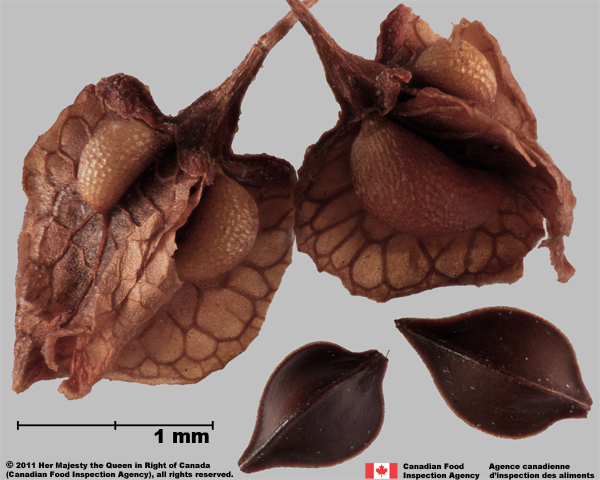
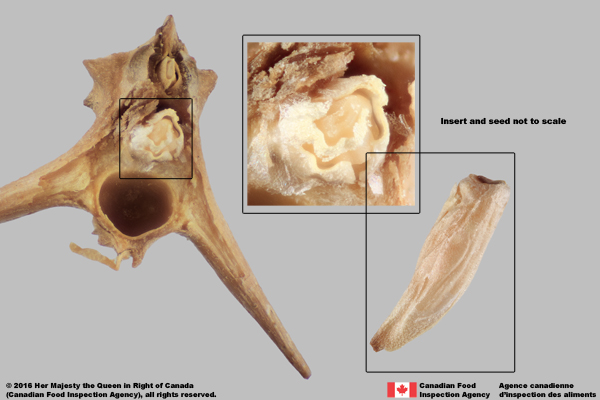
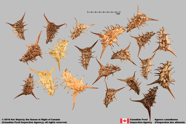
Image Type IV: A group image: to display the potential range of variation in morphological features of a species.
The variation range of size, shape, colour, and dimorphic or heteromorphic seeds, can be captured in a group of 5-10 seeds, or up to 20 seeds if there is a large amount of variation present.
-
The group needs to show the variation in the population and have the seeds in various positions.
-
Arrange the seeds so they are not touching each other, but in a manner in which the seeds can be easily compared with one another.
|
|
|
|
|
| Random display of Centaurea virgata subsp. squarrosa (Asteraceae) achenes | Arranged display of Picris echioides (Asteraceae) achenes |
|
|
|
|
|
|
|
Centaurea solstitialis (Asteraceae) achenes from inner florets |
Centaurea solstitialis (Asteraceae) achenes from outer florets |
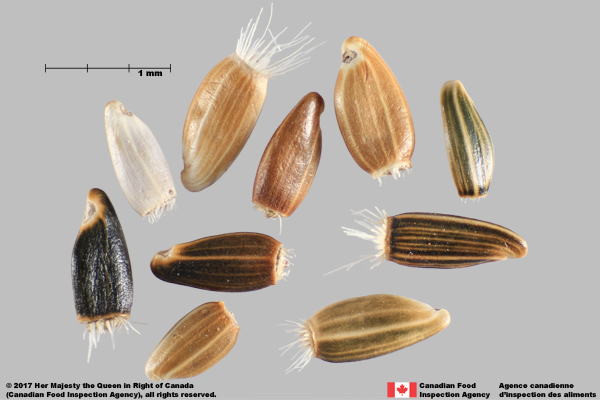
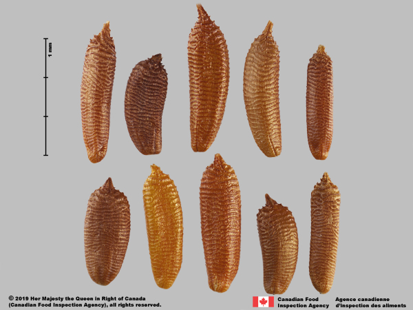
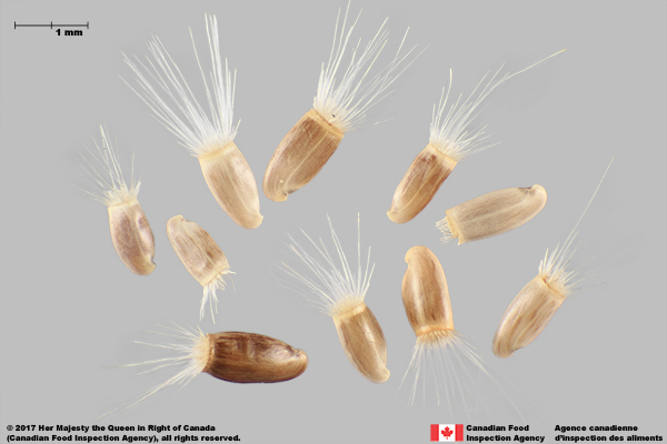
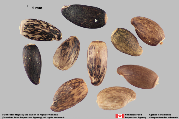
Image Editing and Labelling:
Image standard background:
-
Imaging is recommended to have a pre-determined background colour, such as grey, black or white.
-
Certain features may need additional edits to display the feature properly, such as a white pappus display, if necessary.
-
Image software can calibrate to a standard colour, increasing the consistency of the image background.
The scale must be labeled accurately and clearly for end-users:
-
A physical scale, calibrated ruler, or microscope ruler must be used in imaging
-
Reference scale and equipment scale must be calibrated using known standards or a calibration ruler.
-
International standard length units must be used, such as 0.5 mm, 1.0 mm, 1.0 cm.
Botanical terms or particular seed features shall be labeled, where there is a need and appropriate.
Copyright statement can be marked on the final version for publication.
For examples, image of false wild oat (Avena sativa mut. fatuoid) florets with a standard grey background, scale labeled with the unit, botanical terms specified, and copyright statement
|
Florets false wild oat (Avena sativa mut. fatuoid) |

Image Size for ISMA Publication:
-
It is recommended that an image file be saved in TIFF file format for editorial and storage purposes. For publication purposes, ISMA accepts only JPEG, PNG, and GIF file formats. For more details regarding the different file formats please refer to 'What's the Difference Between PNG, JPEG, GIF, and TIFF?'
-
A recommended size of an image is approximately 1000 pixels in its longest dimension, but the file size must be below the maximum limit listed on the ISMA website.
-
The final image and its associated information, such as caption, seed shape, size, and colour shall be uploaded to the “Author submission” page of the ISMA website.
Authored by: |
Images by: |
|
|
ISMA Editorial Board for ®Seed Identification Guide |
©2019 Canadian Food Inspection Agency, all right reserved. |
©2019 International Seed Morphology Association, all right reserved.